

Categories
Change Password!
Reset Password!



Proximal fibular osteotomy improves pain, knee function, joint space width, and varus alignment at 12 months in patients with medial compartment knee osteoarthritis.
A minimally invasive surgical technique known as proximal fibular osteotomy (PFO) demonstrated remarkable functional and radiographic benefits in medial compartment knee osteoarthritis (OA), according to a prospective 12-month study conducted in Nepal.
The study enrolled 42 patients with Kellgren-Lawrence grade II-III medial compartment OA who underwent PFO at a tertiary care center. Researchers assessed pain, function, and radiographic changes before surgery and at six- and 12-month follow-up visits. By 12 months, patients experienced remarkable reductions in pain and marked improvements in mobility and daily function following PFO. Radiological evaluation also illustrated notable structural benefits, including increased medial joint space width and improved tibiofemoral alignment (Table 1).
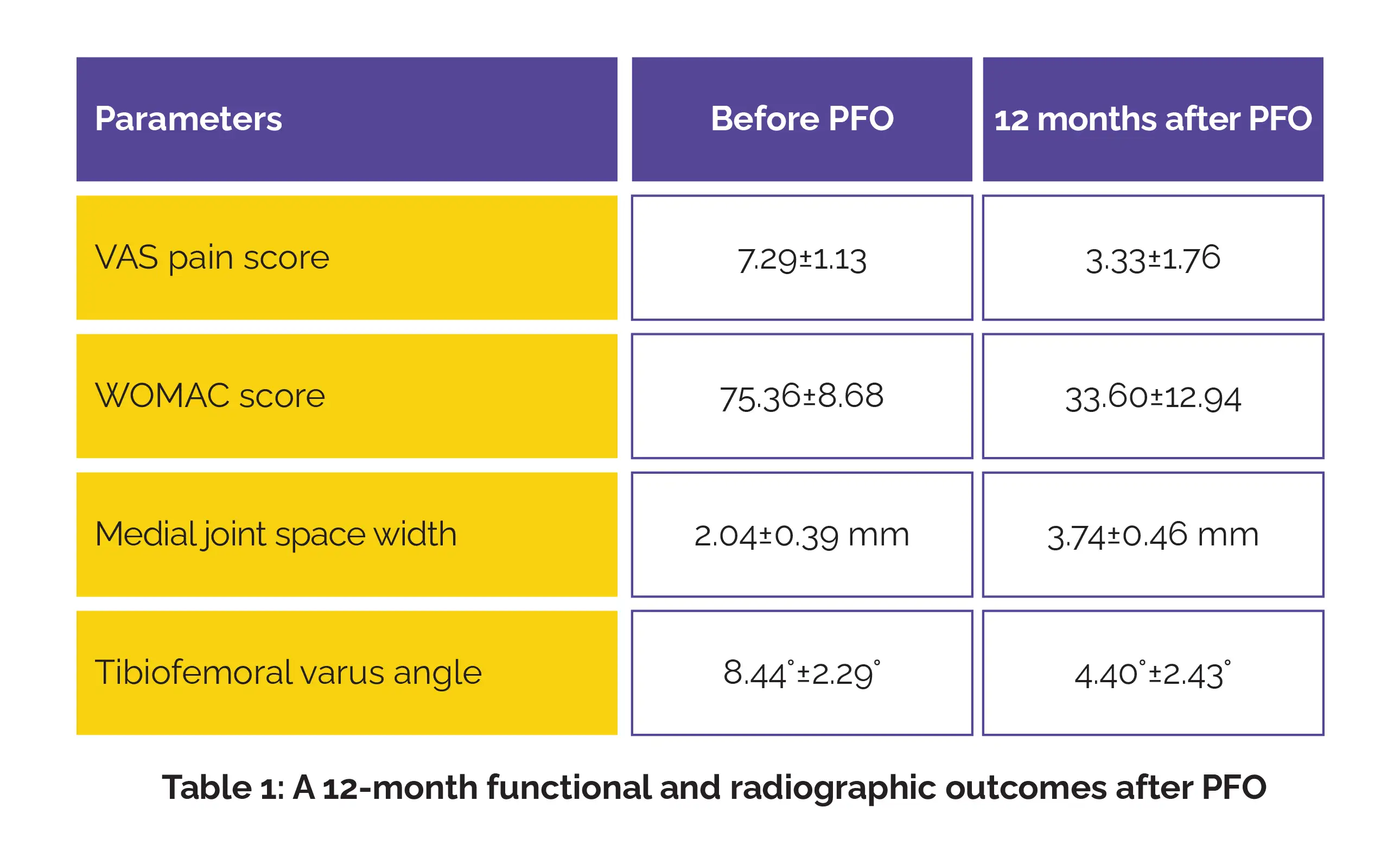
Overall, 61.9% of participants attained excellent functional recovery, defined as at least a 75% reduction in Western Ontario and McMaster Universities Osteoarthritis Index (WOMAC) score. Although the overall complication rate reached 19%, most adverse events were transient and minor.
Investigators concluded that PFO was linked with remarkable reductions in pain, improved knee function, and favorable radiological changes at 12 months, while maintaining an acceptable safety profile. They noted that the procedure may represent a practical and cost-effective joint-preserving option for selected people with medial compartment knee OA, particularly in resource-constrained settings.
Cureus
Functional and Radiographic Outcomes of Proximal Fibular Osteotomy in Medial Compartment Knee Osteoarthritis: A Prospective Single-Arm Interventional Study With 12-Month Follow-Up
Shailesh Dahal et al.












Comments (0)