Категории
Сменить пароль!
Сброс пароля!


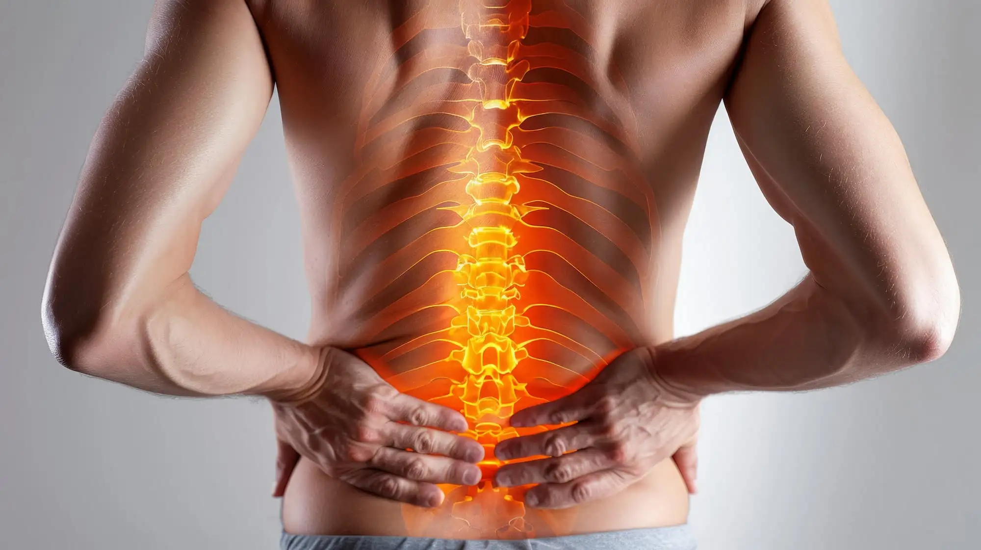
Подагра — распространенное метаболическое заболевание, поражающее периферические суставы. Поражение суставов аксиального скелета встречается редко и часто остается недиагностированным. У 27-летнего мужчины с ожирением и анамнезом плохо контролируемой подагры в течение ≥ 10 лет развилась постоянная боль в области поясницы с левой стороны. При магнитно-резонансной томографии (МРТ) выявлено усиление сигнала на T1-взвешенных изображениях и эрозия по типу «пробойника» в дугоотростчатом суставе L4-L5, что соответствует подагрической артропатии дугоотростчатого сустава.
Данный случай подчеркивает важность включения подагрического спондилита в дифференциальный диагноз у пациентов с хронической болью в спине и длительно существующей гиперурикемией. Ранняя диагностика имеет решающее значение, поскольку тофусные отложения могут вызывать сдавление нервов, нестабильность и хроническую боль. Этот случай подчеркивает необходимость осведомленности врачей об атипичных проявлениях подагры для предотвращения необратимых осложнений со стороны позвоночника и улучшения функциональных результатов.
Общая информация
Подагра — метаболическое заболевание, характеризующееся накоплением кристаллов натрия моноурата в мягких и суставных тканях, что приводит к воспалению и структурным нарушениям. Хотя чаще всего поражаются периферические суставы, у пациентов с хроническим или плохо контролируемым заболеванием все чаще выявляется поражение позвоночника. Подагра пояснично-крестцового отдела позвоночника может сопровождаться хронической болью в спине, радикулопатией и функциональными нарушениями, часто имитируя распространенные патологии позвоночника. Раннее выявление имеет решающее значение для предотвращения неврологических осложнений и необратимых структурных нарушений.
История заболевания
Обсуждение
Этот случай подчеркивает редкость поражения позвоночника при подагре и важность раннего и непрерывного проведения гипоурикемической терапии для предотвращения атипичных проявлений заболевания. Подагрический спондилит следует подозревать у пациентов с хронической подагрой, ожирением и у лиц мужского пола, предъявляющих жалобы на постоянную боль в спине. Хотя подагрический спондилит обычно встречается у пожилых людей, иногда он может возникать и у молодых людей с длительным или плохо контролируемым заболеванием и дополнительными факторами риска, такими как нарушение функции почек.
Диагностика затруднена из-за совпадения симптомов с более распространенными заболеваниями позвоночника, такими как грыжа межпозвоночного диска или стеноз позвоночного канала. Основными методами диагностики остаются МРТ и гистопатологическое исследование. Однако двухэнергетическая компьютерная томография (ДЭКТ) предлагает надежную, неинвазивную альтернативу с высокой чувствительностью и специфичностью в отношении отложений уратов, несмотря на ее более высокую стоимость и ограниченную чувствительность на ранней стадии. Подагрический спондилит остается недооцененным и недостаточно изученным заболеванием, при этом имеются лишь ограниченные данные о долгосрочных результатах и оптимальном лечении.
Лечение аналогично лечению подагры периферических суставов— острые приступы купируются нестероидными противовоспалительными препаратами (НПВП), колхицином или кортикостероидами, в то время как длительная гипоурикемическая терапия (например, аллопуринолом или фебуксостатом) предотвращает рецидивы. Крайне важно уделять внимание таким изменяемым факторам риска, как ожирение, употребление алкоголя и пищевые привычки.
Для точной диагностики и комплексного лечения рекомендуется междисциплинарный подход с участием специалистов спортивной медицины, ревматологов и специалистов по лечению боли. С точки зрения спортивной медицины этот случай подчеркивает, что не все боли в спине имеют механический характер: в основе симптомов со стороны опорно-двигательного аппарата могут лежать системные нарушения обмена веществ, такие как подагра. Распознавание подагрического спондилита в рамках дифференциальной диагностики у пациентов с известным заболеванием подагрой может способствовать проведению более целенаправленных вмешательств и улучшению результатов лечения.
Значение для обучения
Cureus
Facet Joint Gouty Arthropathy: An Uncommon Cause of Chronic Lumbar Pain
Cara C. Chua и соавт.


Комментарии (0)