Categories
Change Password!
Reset Password!


During the postpartum period, a 19-year-old second-time mother presented with severe back pain and difficulty walking, specifically experiencing pain in the right buttock. Pregnancy- and lactation-associated osteoporosis represented an uncommon source of back pain, with the occurrence of a subchondral fracture of the femoral head being exceedingly rare within this condition. For its timely diagnosis, an augmented level of suspicion is crucial. Failure to promptly diagnose and treat this ailment can trigger severe complications.
During pregnancy, low back pain is commonly encountered and can stem from multiple sources. However, it's crucial to consider PLAO as a rare but significant potential diagnosis. Prompt identification and management are imperative to prevent potential complications like vertebral fractures.
In July 2022, a 19-year-old woman presented with severe back pain and pain in her right hip while in her postpartum period. The onset of symptoms commenced as low back pain during pregnancy's final trimester, which was initially tackled using acetaminophen and rest. However, the pain exacerbated after delivery, extending to her back, right buttock, and thigh, substantially impairing her ability to walk. Sitting or standing became extremely challenging for her. This marked her second pregnancy, with an uneventful antenatal period.
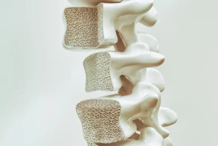
In 1955, pregnancy- and lactation-associated osteoporosis (PLAO) was initially recognized by Nordin and Roper as an infrequent source of severe back pain during the postpartum phase. This ailment is both rare and hazardous, potentially leading to atraumatic fractures during pregnancy and the postpartum period. Its onset typically occurs in the third trimester and persists throughout the postpartum phase. Although the precise pathophysiology remains elusive, various factors such as genetics, race, pregnancy-elicited hypertension, physiological alterations during pregnancy, preterm labor, and obstetric history are implicated as risk factors for PLAO.
One suggested mechanism for PLAO incorporates maternal bone resorption during pregnancy, attributed to insufficient calcium reserves to fulfill the needs of both the mother and the fetus. Furthermore, hormone-triggered demineralization of trabecular bone may occur to supplement calcium for maternal milk production. Pregnancy-related osteoporosis is classified into four types:
During the postpartum and lactating periods, post-pregnancy spinal osteoporosis is an infrequent source of back pain. If left undiagnosed and untreated, it may arouse complications like deformities and vertebral fractures. Given that back pain is a prevalent issue during pregnancy owing to various factors and diagnostic tools like X-rays and bone densitometry are typically avoided during the pregnancy phase, the true incidence of this ailment may be underreported. Fractures impacting the ribs, sacrum, pubic rami, clavicle, and vertebrae have been reported in the literature.
However, no reports of fractures in the subchondral region of the femoral head in lactation-linked osteoporosis have been documented until now. This report elucidates a case of a 19-year-old woman who developed postpartum osteoporosis with fractures in the femoral head, vertebrae, ribs, clavicle, and pelvic rami. It aims to provide a detailed description of the condition known as PLAO in the context of multiple pregnancies.
MEDICAL HISTORY
There were no reports of trauma, infections, or constitutional symptoms throughout both the antenatal and puerperal periods. Additionally, there were no similar occurrences during the patient's first pregnancy.
During examination, the women exhibited a kyphotic posture and an ectomorphic stature. General and systemic assessments did not reveal any specific findings indicative of a diagnosis. No signs of local inflammation or infection were reported in the back. However, tenderness was noted over the dorsolumbar spine and anterior and posterior regions of the right hip. In the sacroiliac joints, no tenderness was observed. Pain was elicited on the right side during both passive and active straight leg raising tests. In the lower limbs, no motor or sensory defects were detected. Examinations for sacroiliac joint and pubic diastasis revealed no abnormalities. Movements of the spine were limited owing to pain, and the patient was unable to independently walk.
The hemogram of the patient displayed normal results, with a hemoglobin level of 11 mg/dL and a total white blood cell count of 4800, comprising 2% monocytes, 3% eosinophils, 45% lymphocytes, and 52% polymorphs. The levels of C-reactive protein and erythrocyte sedimentation rate fell within standard ranges. Results from renal and liver function tests signified normal functioning. The serum levels of parathyroid hormone, vitamin D3, phosphorous, and calcium fell within normal ranges. Serum alkaline phosphatase was measured at 152 IU/L. In the serum electrophoresis, hypergammaglobulinemia was observed, although no M band was detected.
The thoracolumbar spine radiograph illustrated osteopenia accompanied by compression fractures in the L1, D12, and D11 vertebrae. The endplate exhibited normal features, with intact intervertebral disc spaces. Conversely, the pelvis radiograph displayed no abnormalities, with no discernible lesions in the hips or sacroiliac joints. Despite undergoing a skeletal survey, no definitive pathologies were identified. An abdominal and neck ultrasonographic scan was carried out to investigate for potential intra-abdominal, parathyroid, and thyroid irregularities. In the ultrasonographic scan, no abnormalities were identified.
Subsequently, magnetic resonance imaging (MRI) scanning of the spine revealed multiple compression fractures predominantly affecting the dorsolumbar spine. Notably, there was a prominent height decrease in the central portion of the vertebral body, evoking biconcave vertebrae, with the fractures predominantly affecting the D11, D12, and L1 vertebrae. No proof of vertebral body lesions or intervertebral disc space reduction was noted. Additionally, the pelvis MRI examination revealed bone marrow edema impacting the proximal femur.
This was accompanied by a fracture detected in the anterolateral aspect of the femoral head on the right side. On the basis of her clinical presentation, various potential diagnoses were considered, incorporating hyperparathyroidism, tuberculous/pyogenic spondylodiscitis, pubic diastasis, sacroiliitis, intervertebral disc prolapse, and tumors like multiple myeloma and giant cell tumor of the vertebral body. However, subsequent examinations did not support any of these diagnoses.
After ruling out other possible conditions, the diagnosis of PLAO was reached. Treatment involved the administration of vitamin D and calcium supplements along with the utilization of a thoracolumbar brace. Additionally, as part of conservative management, breastfeeding was discontinued. Due to her desire for future pregnancies and the incomplete status of her family, bisphosphonates were not initiated due to their known risks during pregnancy.
Six weeks after beginning treatment, remarkable improvement was noted, with the patient reporting the absence of back or buttock pain. The patient regained the ability to perform all daily activities without any limitations for a period of six months. Presently, the patient continues to be monitored without experiencing any recurrent symptoms.
Postpartum osteoporosis often goes undiagnosed, and its precise incidence remains ambiguous. The mechanisms underlying PLAO remain incompletely understood. Proposed factors contributing to PLAO encompass decreased levels of estrogen and calcium insufficiency during pregnancy and lactation, as well as elevated calcium demands during these periods. Some researchers also suggest a genetic predisposition to the ailment.
Dunne et al. observed a higher prevalence of fractures among mothers of patients suffering from PLAO compared to mothers of controls, indicating a potential genetic influence. The primary symptom frequently observed in postpartum osteoporosis is back pain. Therefore, it's crucial to examine all pregnant and lactating women who exhibit severe back pain for this condition. No specific therapeutic guidelines are currently established for this ailment. Typically, patients are managed with the help of vitamin D and calcium supplements and are motivated to wean.
Certain studies propose that bisphosphonates could be beneficial in treatment. But, there are concerns regarding the potential accumulation of bisphosphonates in maternal bones and the risk of skeletal ailments in subsequent pregnancies. Consequently, these treatments are generally evaded in those planning future pregnancies. Teriparatide (a human recombinant form of parathyroid hormone) proves effective in relieving osteoporotic fractures by enhancing bone mineral density and reducing fracture rates. Unlike bisphosphonates, it doesn't accumulate in the skeleton, making it a safer option for females contemplating future pregnancies.
Teriparatide has exhibited efficacy in enhancing bone mineral density and alleviating clinical symptoms in PLAO sufferers. Patients battling PLAO have a heightened risk of experiencing recurrences in subsequent pregnancies, with some studies indicating a recurrence rate of around 33%. Therefore, it's pivotal to inform patients about the potential for the ailment to recur during future pregnancies.
This case was presented because of the unusual occurrence of a subchondral fracture of the femoral head in PLAO. Formerly, there have been no recorded instances of such fractures connected with PLAO. Diagnosis proved formidable as the patient experienced buttock and back pain without evident hip or sacroiliac joint abnormalities on X-ray. Laboratory tests ruled out common causes of osteoporosis, leading to diagnosis by process of elimination.
Learning
During pregnancy, low back pain is commonly encountered and can stem from multiple sources. However, it's crucial to consider PLAO as a rare but significant potential diagnosis. Prompt identification and management are imperative to prevent potential complications like vertebral fractures.
References
1. Dunne F, Walters B, Marshall T, Heath DA. Pregnancy associated osteoporosis. Clin Endocrinol (Oxf) 1993;39:487- 90.
2. Gandhi S, Rodriguez A, Baim S. MON-505 an atypical case of osteoporosis related to pregnancy and lactation. J Endocr Soc 2019;3:MON-505.
3. Khandelwal D, Tandon N. Overt and subclinical hypothyroidism: Who to treat and how. Drugs 2012;72:17-33.
4. Holmberg-Marttila D, Sievänen H, Tuimala R. Changes in bone mineral density during pregnancy and postpartum: Prospective data on five women. Osteoporos Int 1999;10:41-6.
5. Polat SB, Evranos B, Aydin C, Cuhaci N, Ersoy R, Cakir B. Effective treatment of severe pregnancy and lactation-related osteoporosis with teriparatide: Case report and review of the literature. Gynecol Endocrinol 2015;31:522-5.
6. Clemetson IA, Popp A, Lippuner K, Ballmer F, Anderson SE. Postpartum osteoporosis associated with proximal tibial stress fracture. Skeletal Radiol 2004;33:96-8.
7. Ofluoglu O, Ofluoglu D. A case report: Pregnancy-induced severe osteoporosis with eight vertebral fractures. Rheumatol Int 2008;29:197-201.
8. O’Sullivan SM, Grey AB, Singh R, Reid IR. Bisphosphonates in pregnancy and lactation-associated osteoporosis. Osteoporos Int 2006;17:1008-12.
9. Lampropoulou-Adamidou K, Trovas G, Stathopoulos IP, Papaioannou NA. Case report: Teriparatide treatment in a case of severe pregnancy-and lactation-associated osteoporosis. Hormones (Athens) 2012;11:495-500.
10. Di Gregorio S, Danilowicz K, Rubin Z, Mautalen C. Osteoporosis with vertebral fractures associated with pregnancy and lactation. Nutrition 2000;16:1052-5.


Comments (0)