Categories
Change Password!
Reset Password!


With age, pelvic diameters shrink, iliac spine distance and subpubic angle widen, and a previously undocumented downward-extending sciatic notch spine appears in 25% of women, potentially affecting hip health.
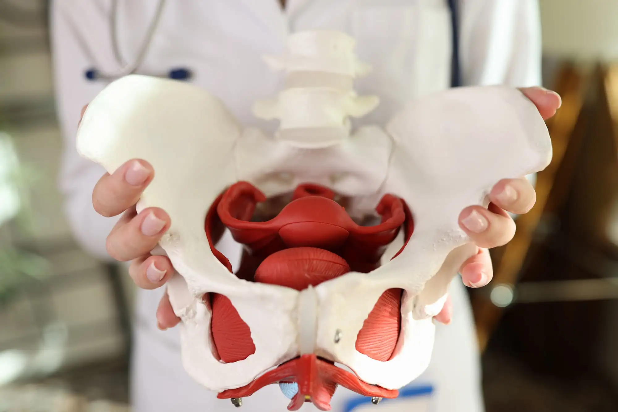
A comprehensive radiological analysis uncovers age-linked shifts in pelvic dimensions among Turkish women, revealing a newly identified anatomical feature with potential clinical relevance. The study aimed to characterize pelvic morphometry in adult women and to determine how these measurements varied across age groups.
The research team analyzed computed tomography (CT) scans from 324 healthy women aged 18 to 60 years. Investigators measured multiple pelvic parameters, including anatomical, obstetric, and diagonal conjugates, iliac dimensions, subpubic angles, pelvic inclinations, and structural alignments. Age groups were stratified, and morphometric variables were compared using statistical methods to identify correlations and group-based differences.
The study also evaluated potential lateral asymmetries and documented any unusual anatomical features that appeared consistently across the sample. The analysis showed a significant age-related decline in the anatomical, obstetric, and diagonal conjugate diameters, with strong negative correlations across all three measures. Differences among age categories were confirmed as statistically significant. No lateralization was detected, except for the articulation pattern within the pubic symphysis.
The subpubic angle varied markedly with age, demonstrating significant group differences. An inter-anterior superior iliac spine distance increased with age and also differed markedly across age groups. Pelvic inclination at both the inlet and outlet displayed meaningful variation with advancing age. Unexpectedly, approximately one-quarter (25%) of participants presented a downward-projecting spinous process originating from the superior border of the greater sciatic notch.
Researchers noted that this projection could impose pressure on adjacent neurovascular structures. The study demonstrated distinct age-related alterations in pelvic dimensions among Turkish women and identified a previously unreported anatomical variation that may contribute to hip discomfort or neurovascular compression.
Bratislava Medical Journal
Morphometric Analysis of Female Pelvic Cavity Among Turkish Population: A Radiological Study
Mustafa Canbolat et al.


Comments (0)