Categories
Change Password!
Reset Password!


Segmental testicular infarction (STI) is a rare, idiopathic condition that poses diagnostic challenges. This report highlights a 38-year-old man with bilateral STI potentially linked to iron deficiency anemia (IDA). The patient presented with chronic bilateral testicular pain, more pronounced on the left side. Laboratory tests revealed elevated white blood cells, monocytes, neutrophils, C-reactive protein, and platelet count, alongside decreased hemoglobin and red cell indices.
Imaging studies, including ultrasound and MRI, identified hypoechoic, avascular areas in both testes. Conservative treatment with anti-inflammatories, antibiotics, anticoagulants, and iron supplementation led to significant improvement. Follow-up ultrasounds over a month revealed complete healing of lesions without recurrence. This study emphasizes the importance of considering IDA in the differential diagnosis of STI and highlights the efficacy of non-surgical management.
A 38-year old man presented with:
STI is a rare and idiopathic condition of the testes, with fewer than a hundred cases described in the literature to date. In most instances, the underlying cause remains unclear. However, recent studies have implicated numerous hematologic ailments—such as protein C deficiency, sickle cell anemia, and arterial atheroma—as potential contributors to its pathogenesis. IDA, the most widespread form of anemia globally, is marked by microcytic hypochromic anemia due to depleted iron stores. Beyond its classic manifestations of reduced hemoglobin and erythrocyte levels, IDA is also frequently linked with reactive thrombocytosis, which may predispose the affected individuals to thromboembolic events.
Medical History
(a) Physical
(b) Laboratory
(c) Imaging
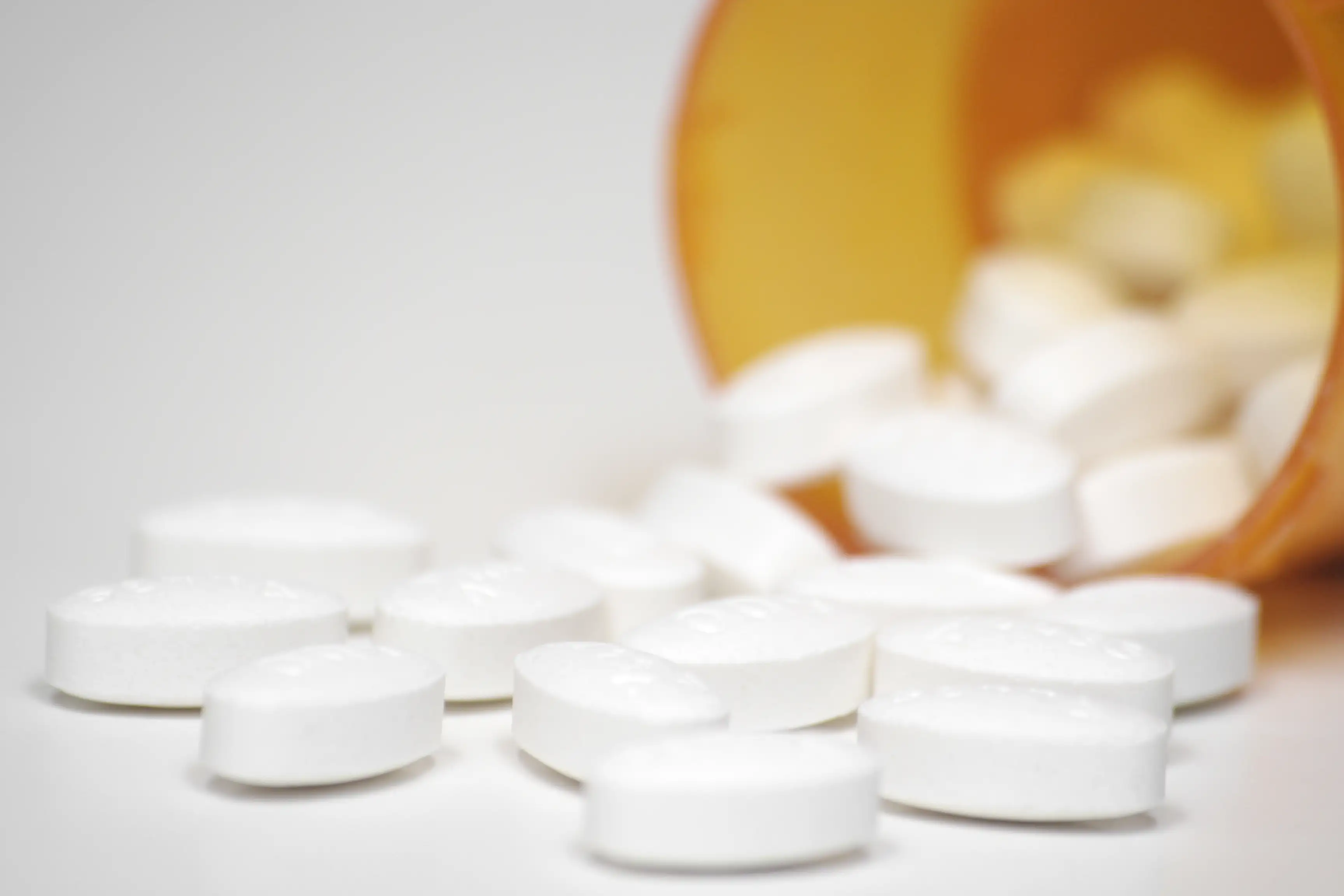
Initial management included anti-inflammatory and antibiotic therapy, along with rivaroxaban for anticoagulation. Upon identification of severe iron deficiency, the patient was started on daily intravenous iron sucrose. He continued iron supplementation after discharge. After 3 days of treatment, the patient experienced slight relief in testicular pain. Inflammatory markers showed a decrease, although platelet counts remained elevated.
Following treatment for another 3 days, there were substantial increases in ferritin and hemoglobin levels. This was accompanied by decreases in platelet counts and CRP levels, along with marked relief in testicular pain. Ultrasound imaging revealed a prominent reduction in the hypoechoic areas in both testes.
Two weeks after discharge, ferritin and hemoglobin levels had normalized, and platelet counts returned to normal. Ultrasound examination showed complete resolution of the hypoechoic area in the right testicle and a further reduction in the left. One month after discharge, ultrasound indicated complete resolution of the hypoechoic areas in both testes.
Discussion
STI, a form of ischemic necrosis impacting part of the testicular parenchyma, is notably rarer than many other testicular pathologies. Although the precise mechanisms behind STI remain elusive, emerging evidence has pointed towards a possible vascular origin. Several hematologic ailments—such as arterial atheroma, cholesterol embolism, sickle cell anemia, and protein C deficiency—have been implicated in previous reports, with embolic phenomena suspected as potential triggers.
Notably, 1 large cohort study involving 36,327 IDA sufferers revealed that 32.6% presented with thrombocytosis, and among them, 15.8% experienced thrombotic events. Thromboses linked to IDA have been reported to manifest in diverse clinical forms, including cerebral infarction and central retinal vein occlusion. In the present case, the patient’s longstanding IDA, coupled with marked thrombocytosis, likely created a hypercoagulable environment conducive to vascular occlusion within the testes. Though no pathological specimen was available due to the absence of surgery, clinical improvement following iron supplementation was striking—both in terms of minimized infarct size and relief of bilateral testicular pain. This response adds weight to previous observations where IDA-linked thrombosis resolved with iron therapy and anticoagulation. Taken together, these findings lend further support to the hypothesis that STI, at least in some cases, may represent a vascular complication arising from IDA.
Distinguishing STIs from testicular neoplasms or infections can be clinically challenging. However, in this case, STI diagnosis was relatively straightforward. Unlike testicular tumors, which typically present unilaterally, the patient exhibited bilateral infarctions—an uncommon feature in neoplastic conditions. Color Doppler ultrasound further supported the diagnosis, showing an absence of blood flow in both testes. In contrast, testicular tumors usually illustrate either normal or increased vascularity; while avascular tumors do exist, a complete bilateral absence of blood flow has not been documented in the literature.
Testicular infection was also deemed unlikely. Ultrasound findings in orchitis typically reveal testicular enlargement, heterogeneous echotexture, and increased parenchymal blood flow on Doppler imaging—none of which were present in this case. Moreover, MRI findings provided additional diagnostic clarity. The lesions displayed low signal intensity on T2-weighted images, which contrasts with the high T2 signal commonly seen in both testicular tumors and infections. Negative tumor marker results further ruled out malignancy, collectively reinforcing the diagnosis of bilateral STI.
STI poses a diagnostic obstacle for clinicians owing to its rarity and ambiguous clinical presentation. This report described an uncommon case of bilateral STI likely linked to IDA. With prompt conservative care and iron supplementation, both the patient's hematologic parameters and testicular symptoms improved remarkably. At 1-month follow-up, imaging confirmed total lesion healing, with no signs of recurrence. However, a key limitation of this case lies in the absence of pathological confirmation, as the diagnosis was based solely on imaging findings and clinical progression.
Learning
BMC Urology
Bilateral segmental testicular infarction secondary to iron deficiency anemia: a case report
Yun Duan et al.


Comments (0)